| |
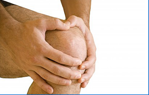
Articular Cartilage Damage
Articular cartilage is the smooth, shiny white lining that covers the ends of the bones inside the knee joint. It allows the joint surfaces to glide smoothly with very little friction, and it also helps to cushion the knee and distribute load across the joint. Unfortunately, articular cartilage has a very limited ability to heal itself. When it is damaged, the body may fill the defect with scar-like repair tissue, but this is not as durable or as smooth as the original cartilage.
Articular cartilage damage may occur after an acute injury such as a twist, fall, direct impact, or sporting injury. This is different from the gradual wear and tear seen in osteoarthritis, and it is important to distinguish between the two because the treatment options and expected outcome can be different. Patients may complain of pain, swelling, catching, clicking, giving way, or occasional locking of the knee.
Diagnosis
The diagnosis of articular cartilage damage can sometimes be difficult. Standard X-rays do not show cartilage directly, but they are useful for assessing the overall condition of the knee and for identifying other problems. MRI scans can be helpful in detecting cartilage injury and in looking for associated problems such as meniscal tears, ligament injuries, or bone bruising. In some cases, the most accurate way to assess the size, site, and depth of the cartilage defect is by knee arthroscopy, which allows direct inspection of the joint surface.
Treatment
Treatment depends on a number of factors, including the size and location of the defect, the condition of the underlying bone, any associated injuries, and the patient’s age, symptoms, and activity level. Not every cartilage lesion requires the same treatment. In selected cases, surgery may be recommended to improve symptoms, restore function, and help protect the joint in the longer term.
1. Arthroscopic Debridement / Chondroplasty
This is a keyhole procedure and is usually performed as a day-case operation. The knee is inspected arthroscopically and the damaged cartilage is carefully trimmed and smoothed, removing any loose or unstable flaps while preserving the healthy surrounding cartilage. This procedure is mainly used in selected milder lesions, particularly where the damaged cartilage is causing mechanical symptoms such as catching or irritation within the joint. Recovery is usually quicker than with cartilage restoration procedures, and many patients return to normal day-to-day activities relatively soon afterwards.
2. Arthroscopic Microfracture
This is also a keyhole procedure and is used for selected full-thickness cartilage defects where the bone beneath the cartilage is exposed. After the damaged area has been cleaned and prepared, a number of small holes are made in the underlying bone to encourage a healing response. This allows repair tissue to form within the defect. The new tissue is fibrocartilage rather than normal articular cartilage, so microfracture is generally better suited to smaller, well-contained lesions in carefully selected patients. Following surgery, a period of protected weight-bearing with crutches and a structured physiotherapy programme is often required to give the repair tissue the best chance to develop.
In some patients, depending on the size and site of the defect, other cartilage restoration procedures may also be considered. Current guidance and recent reviews note that larger symptomatic defects may be better suited to other restoration techniques rather than microfracture alone.
Further detailed information and the appropriate treatment option will be provided to you by Mr Ayoub during the consultation. Back...
|
|